

Mission
Read and understand complex medical phenomena and work with multidisciplinary teams of doctors to produce images and animations which are both scientifically rigorous and didactically clear.
My role: Visual Designer, Motion Designer, Concept Illustrator.
Tools: Illustrator, Photoshop, Corel Painter, After Effects, reading.
The landscape: Medicine is a complex field which requires excellence, peoples' lives depend on it. Doctors and medical students must wrap their heads around intricate anatomic and physiologic concepts in order to deliver accurate diagnoses for their patients. The illustrations and animations that I make for them must be painstakingly accurate and convey subtle information clearly.
Clients
Department of Defense Joint Pathology Center
Walter Reed National Military Medical Center
American Institute of Radiologic Pathology, Radiologic Residency Program
American College of Radiology
University of Maryland Medical School
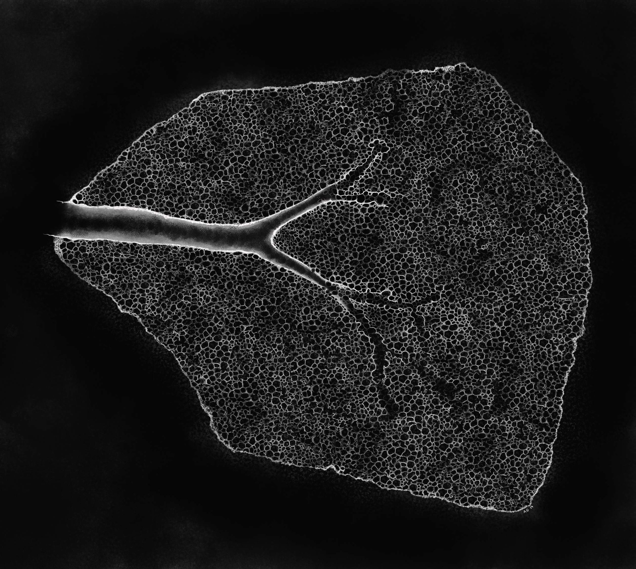
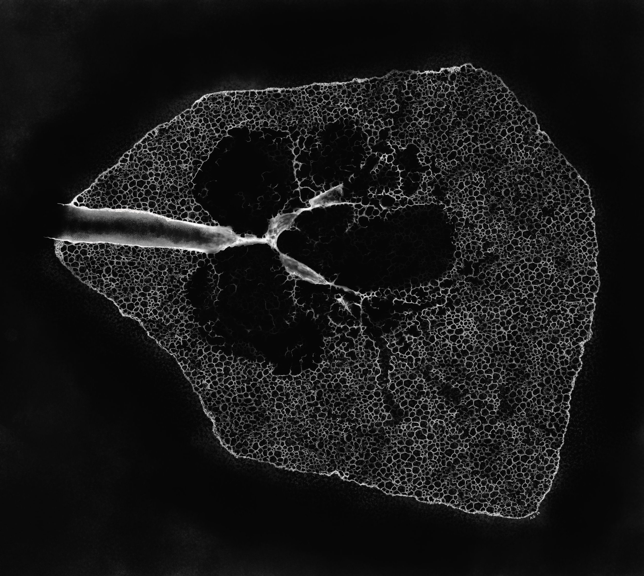
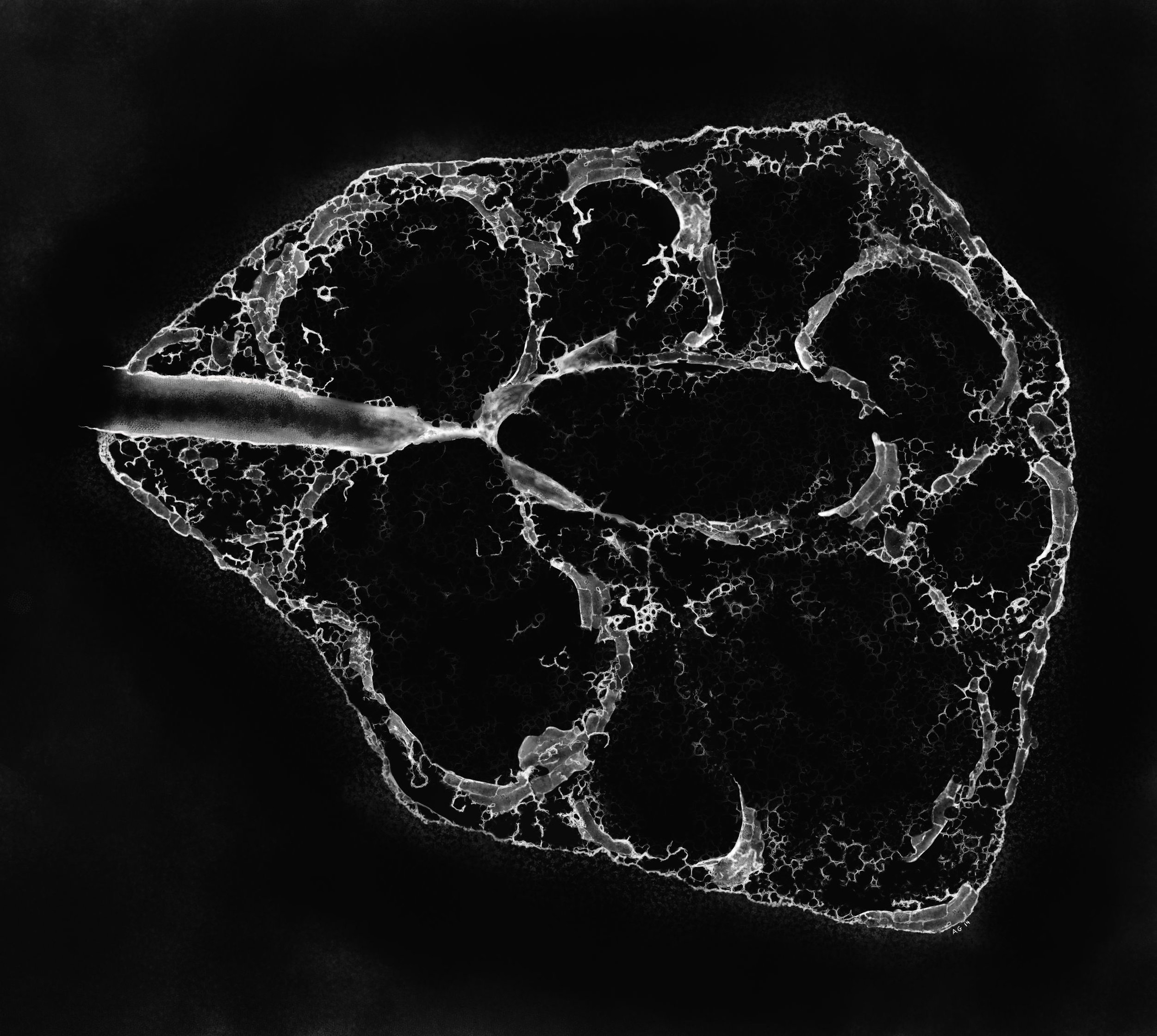
01 // Emphysema
These illustrations were commissioned for use in post graduate education lectures at the American College of Radiology and the American Institute of Radiologic Pathology. They are freehand digital drawings of sequential sections of human lung which demonstrate the effects of emphysema, and their underlying cause: particulate matter from cigarette smoking.
These detailed visualizations required an in-depth study of Dr. Ewald Weibel's landmark works on lung structure and function, lung specialists at the American Institute of Radiologic Pathology guided me through this research.

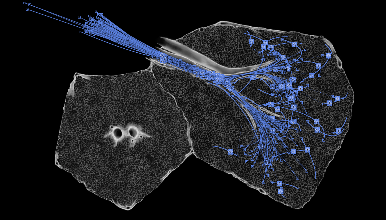
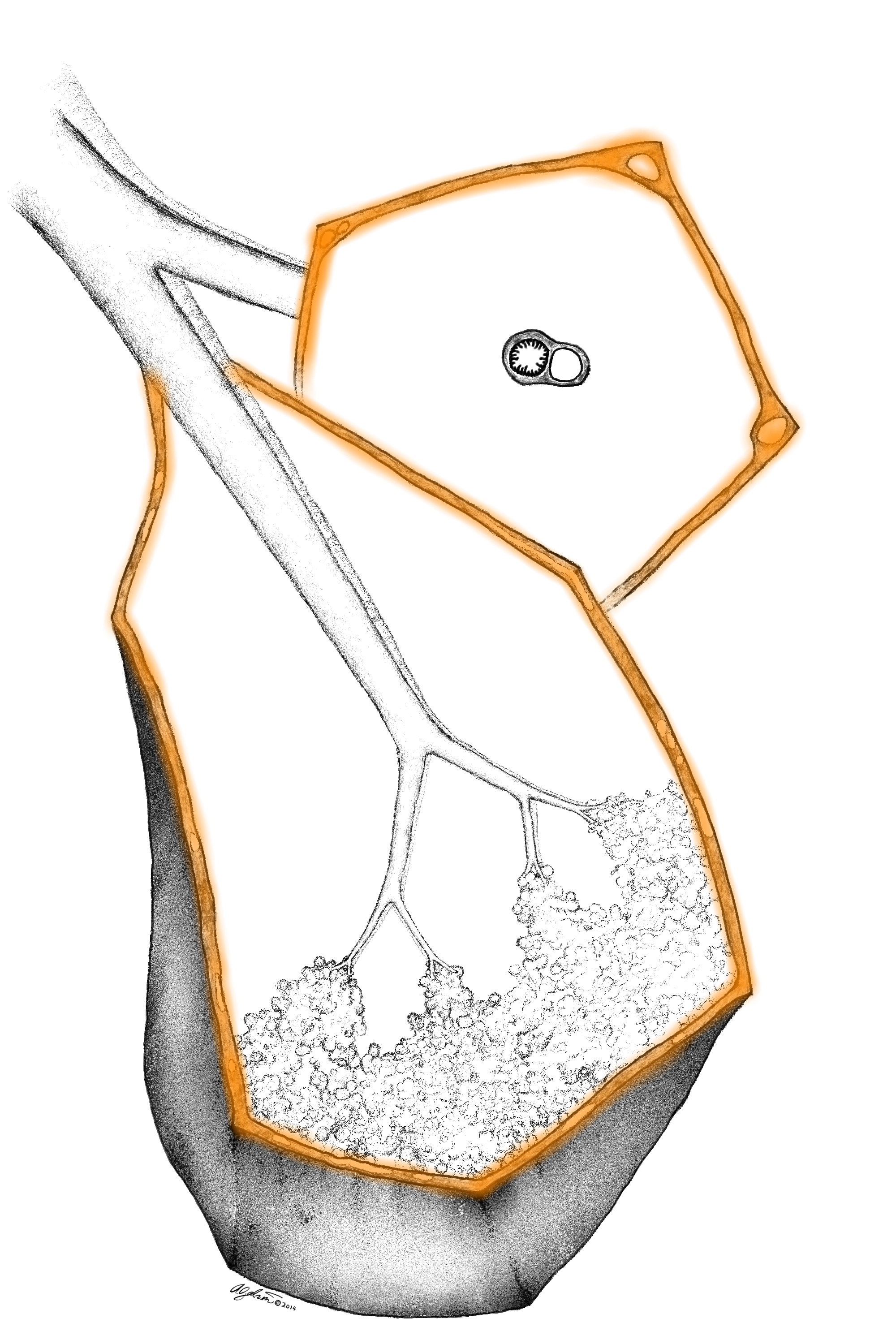
02 // Particle Deposition and Transport
The first phase of this animation shows harmful particles which are carried into the secondary lobule upon inhalation and their classic distribution throughout the lobule. The second phase shows the body's clearance mechanisms at work. Most particles are removed up the conducting airways by the mucociliary elevator, but a few particles are swept out to the inter-lobular septa. As particles are taken up by the lymphatic system, they become yellow.


03 // Gravity Based Alveolar Differential
The anatomy and physiology of the human lung is greatly influenced by gravity. Because the alveoli of the lung are actually suspended from a fine web of connective tissue, at rest the alveoli at the lung apex are stretched to their maximum capacity, whereas alveoli at the lung base are flattened. This is a critical piece of information because this differential affects which regions of the lung are most heavily supplied with blood, air and lymphatic flow.
04 // Pulmonary Hydrostatic Pressure
The upright human lung sits in a gravitational field. Not only does this affect its structure, but dictates the movement patterns of blood, air and lymph. Blood, traveling from the pulmonary arteries into the parenchyma for oxygenation does not flow equally into all parts of lung. Blood vessel pressure is greater in the lung base than it is at the apex.
Dr. John West's classic zones of perfusion and their anatomic location are represented by grey strokes.
Zone 1 alveolar pressure > arterial pressure > venous pressure
Zone 2 arterial pressure > alveolar pressure > venous pressure
Zone 3 arterial pressure > venous pressure > alveolar pressure
Zone 4 the weight of the lung itself presses on the blood vessels, constricting their diameter slightly such that blood flow is actually slightly less


05 // Lymphatic Drainage
Because blood flow is greater to the lung bases, lymphatic clearance is greater in the lung bases as well. Hydrostatic pressure drives the movement of fluid and small molecules into the lymphatic vessels which pervade the interlobular septa of the lung. This means that foreign particles and damaging substances are more likely to be removed from the lung base.

06 // Secondary Lobule Distribution Patterns
The secondary lobule is a fractal microcosm of the whole lung. Disease processes occur in different locations in this structure, which lead to visually distinct distributions.

Septal pattern : human lung tissue
Septal pattern : illustration